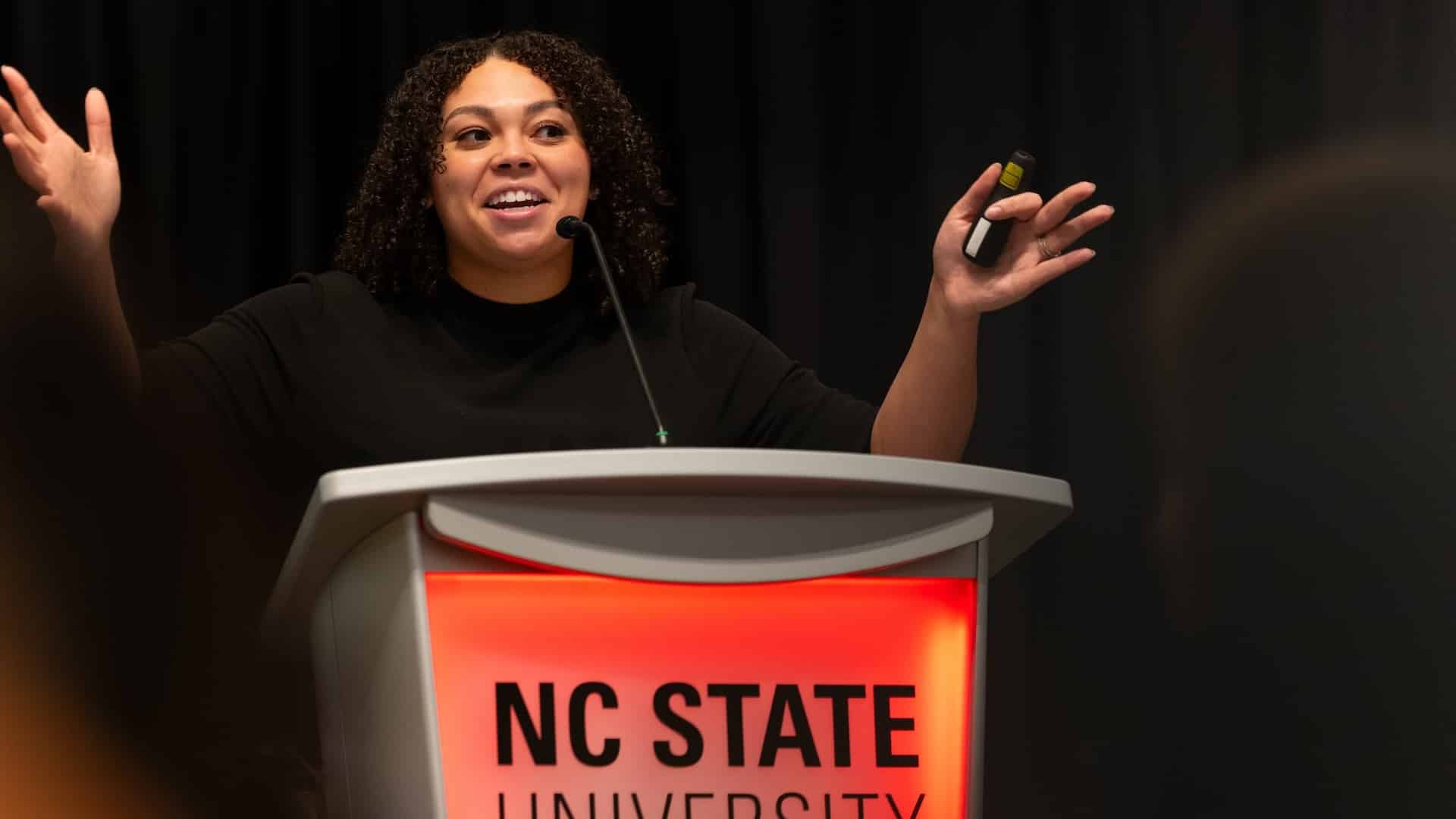
A Summer of Hands-on Science
Undergraduate students from all over the U.S. spent their summer conducting research in NC State's labs, alongside our faculty and graduate students.
Classes are officially back in session and the Wolfpack is settling into a new semester. Before summer slips away from us, we’re looking back at the work our faculty, staff and visiting students accomplished over the break.
Through a number of National Science Foundation-funded Research Experiences for Undergraduates (REU) programs, we welcomed students from other universities across the country into our labs and facilities. These students worked alongside our faculty and graduate students, gaining hands-on research experience.
Stefanie Chen, assistant teaching professor of biological sciences, and Melissa Srougi, associate teaching professor of molecular biomedical sciences in the College of Veterinary Medicine — both faculty in the Biotechnology Program — hosted the first Modeling the Dynamics of Biological Sciences REU, or the BioDynamics REU program, which will run for three years.
Take a sneak peek at the group’s summer at NC State.
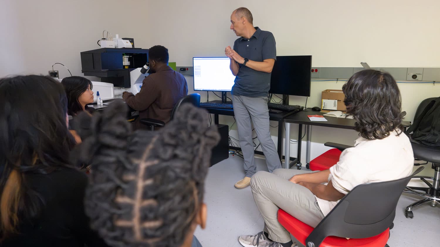
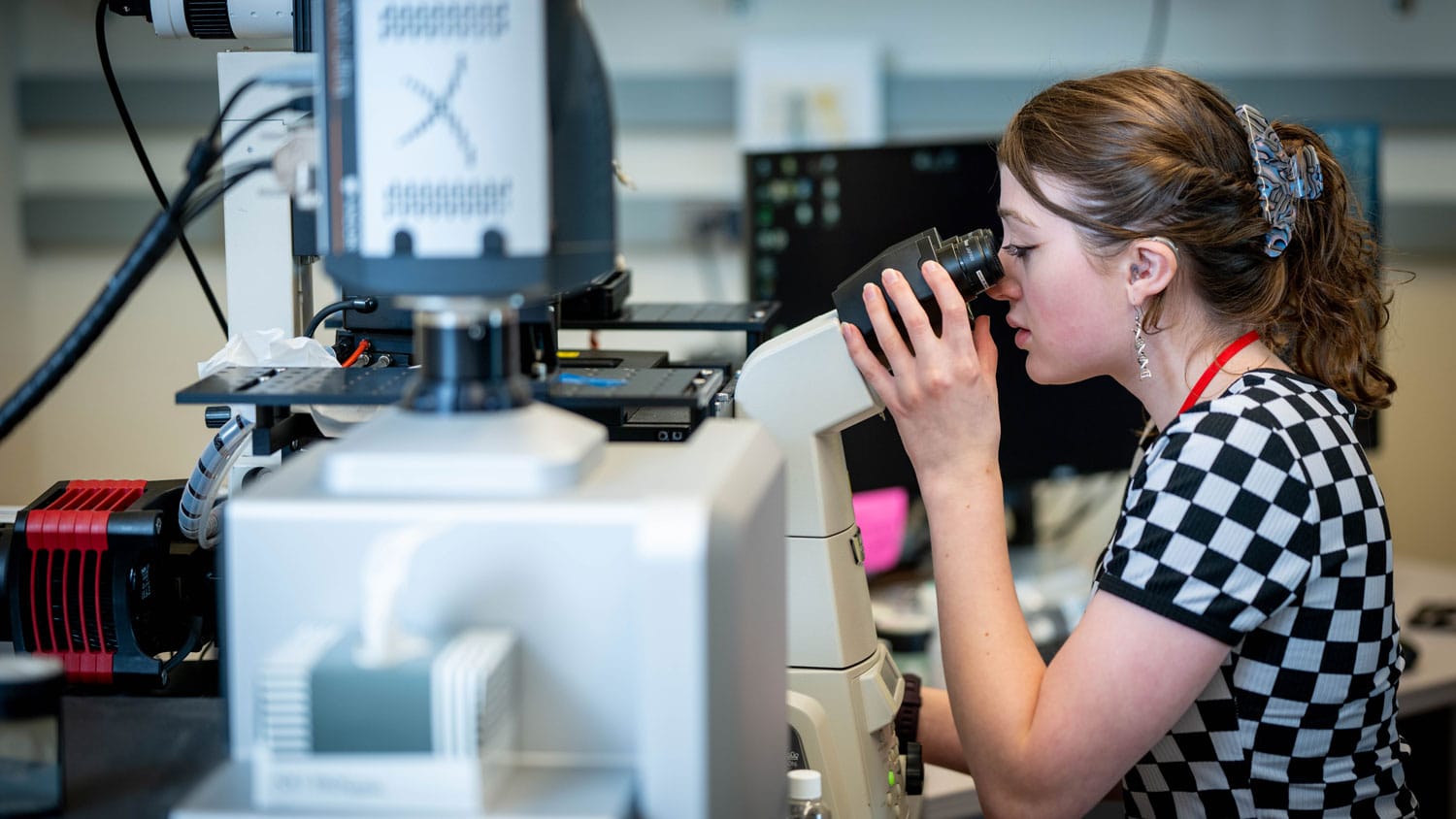
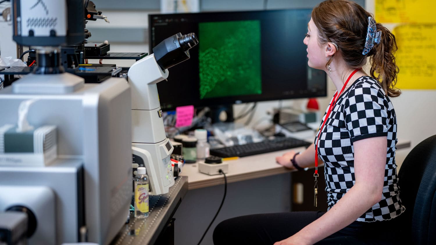
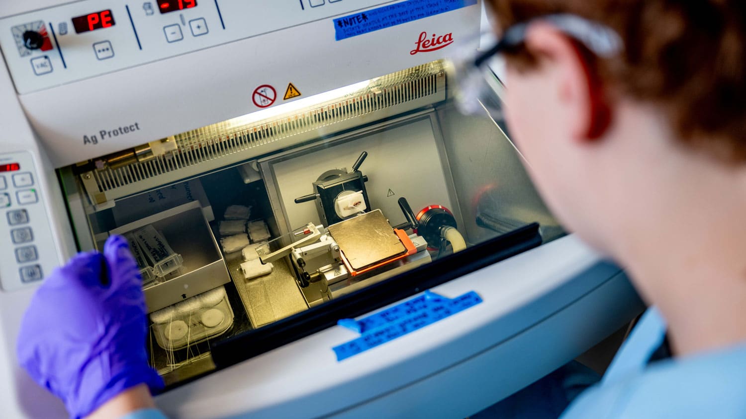
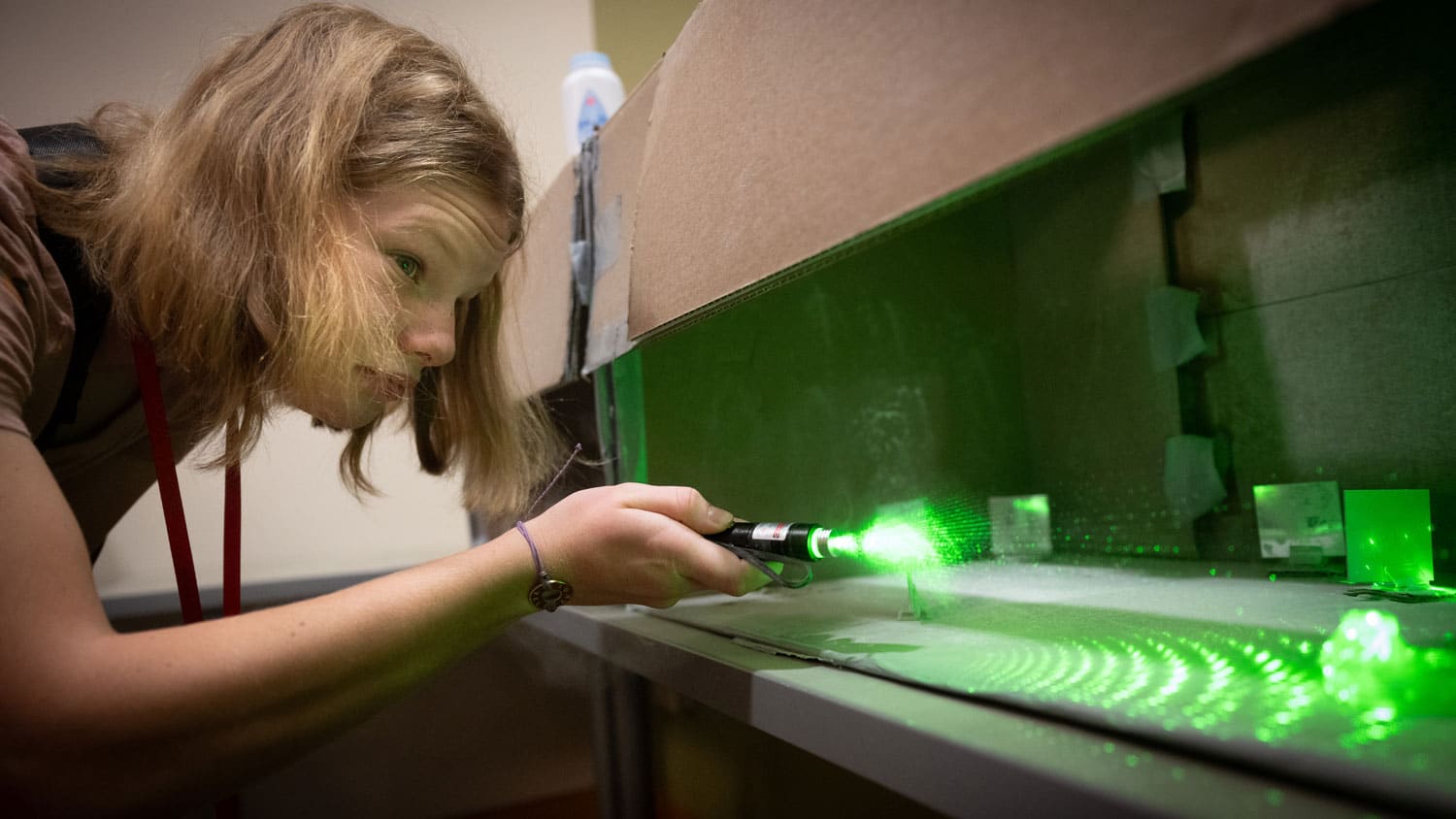
They Took Lessons in Microscopy
Some of the BioDynamics REU students had no prior lab experience. To prepare for 10 weeks of full-time lab work, they spent their first week learning the basics. They took an introductory lecture on light microscopy — a technique to make small objects visible using visible light and lenses — and learned to use electron microscopes and confocal microscopes.
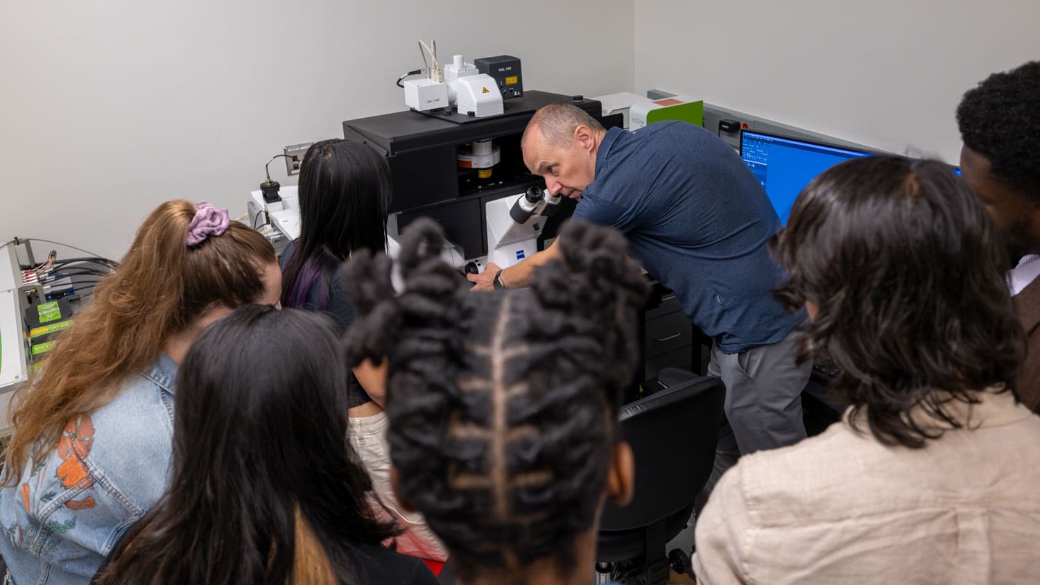
Below, Mariusz Zareba, director of the Cellular and Molecular Imaging Facility in the Plant Sciences Building, shows the group how to use a confocal microscope and analyze the images it produces.
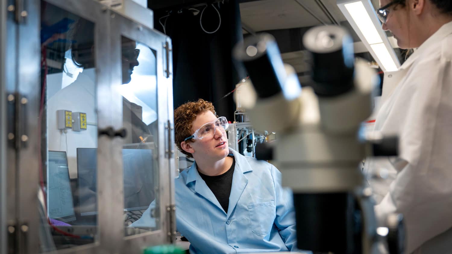
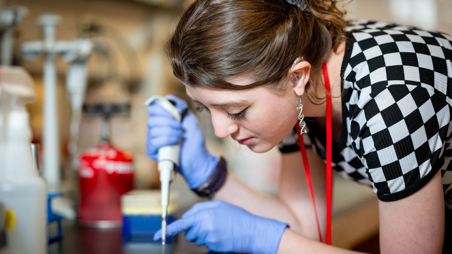
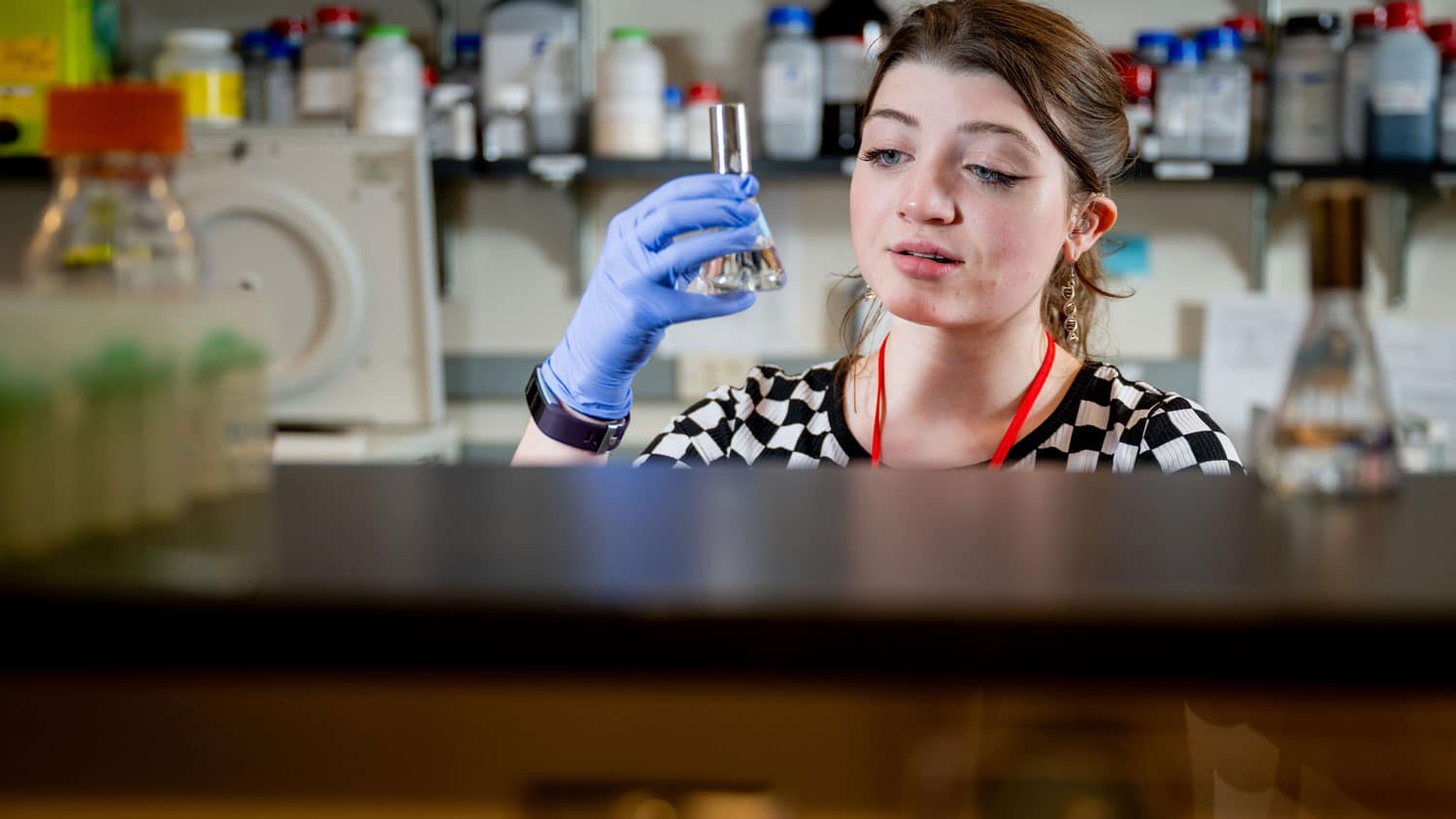
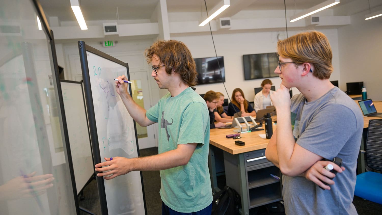
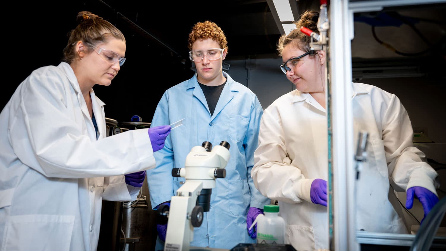
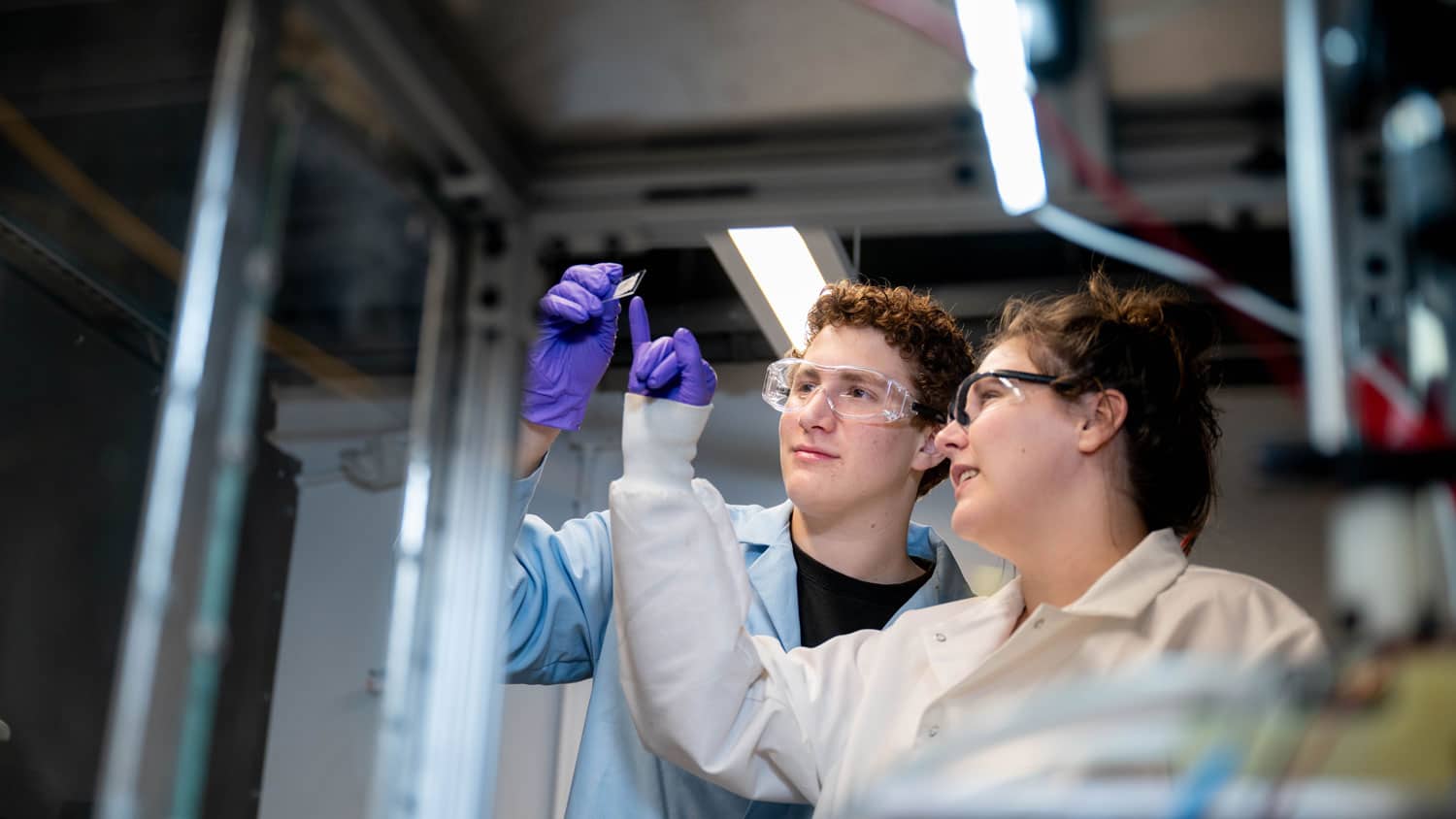
They Were Full-time Scientists
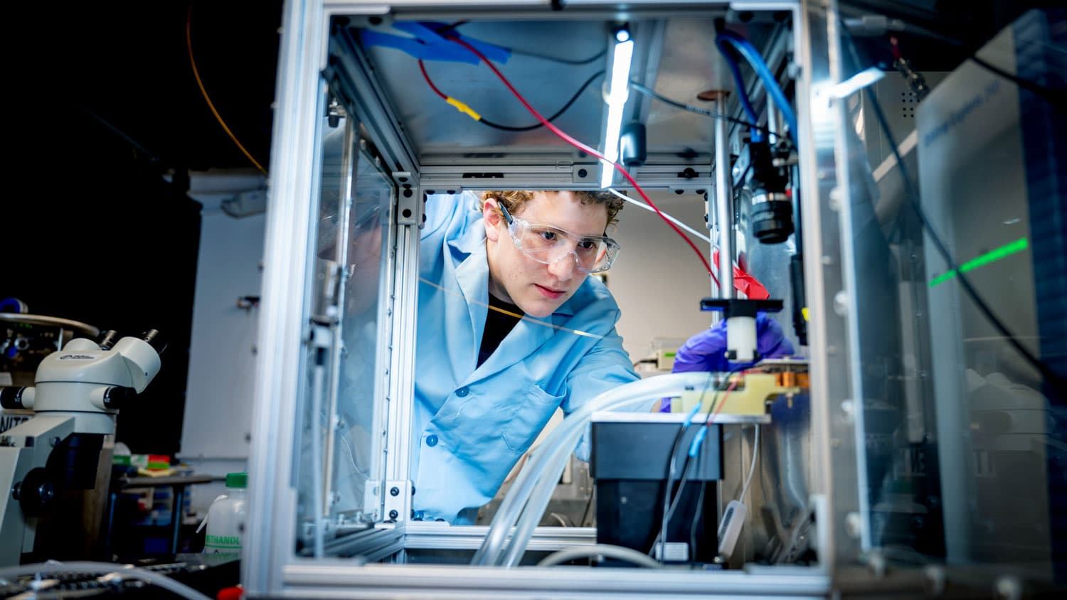
Equipped with the knowledge they needed, the students hit the ground running. They spent their summer days conducting research in labs at the College of Sciences, the College of Veterinary Medicine and the Joint Department of Biomedical Engineering.
We dropped into a few of the College of Sciences labs throughout the summer to get a firsthand look at their research.
Meet Kaelyn Coates, a sophomore majoring in biomedical sciences at the Rochester Institute of Technology in New York state. She worked in the lab of Mary Elting, associate professor of physics, studying the intricacies of cell division, specifically how the mitotic spindle — or the cellular machine that segregates chromosomes when cells divide — carries out its function. The mitotic spindle’s job is critical given that mistakes in cell division can lead to cancer, birth defects and miscarriage.
Coates worked on a variety of projects, including looking at the effects of temperature on spindle elongation and the effects of sorbitol, a sugar alcohol, on osmotic pressure on the nucleus. She also learned the Python programming language, which allowed her to see the connection between science and data analysis more clearly.
Coates was one of three deaf and hard of hearing students in the BioDynamics REU program, which prioritized recruiting from Deaf-serving institutions.
“It was nice to still be able to communicate with people with [American Sign Language], and that others were also willing to learn it,” said Coates. “I had one roommate who was deaf in one ear and hard of hearing in the other, and another roommate who was all hearing, and she learned some ASL so she could communicate with us.”
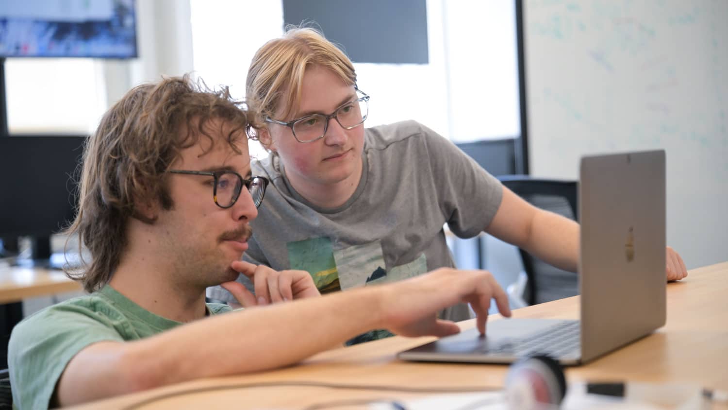
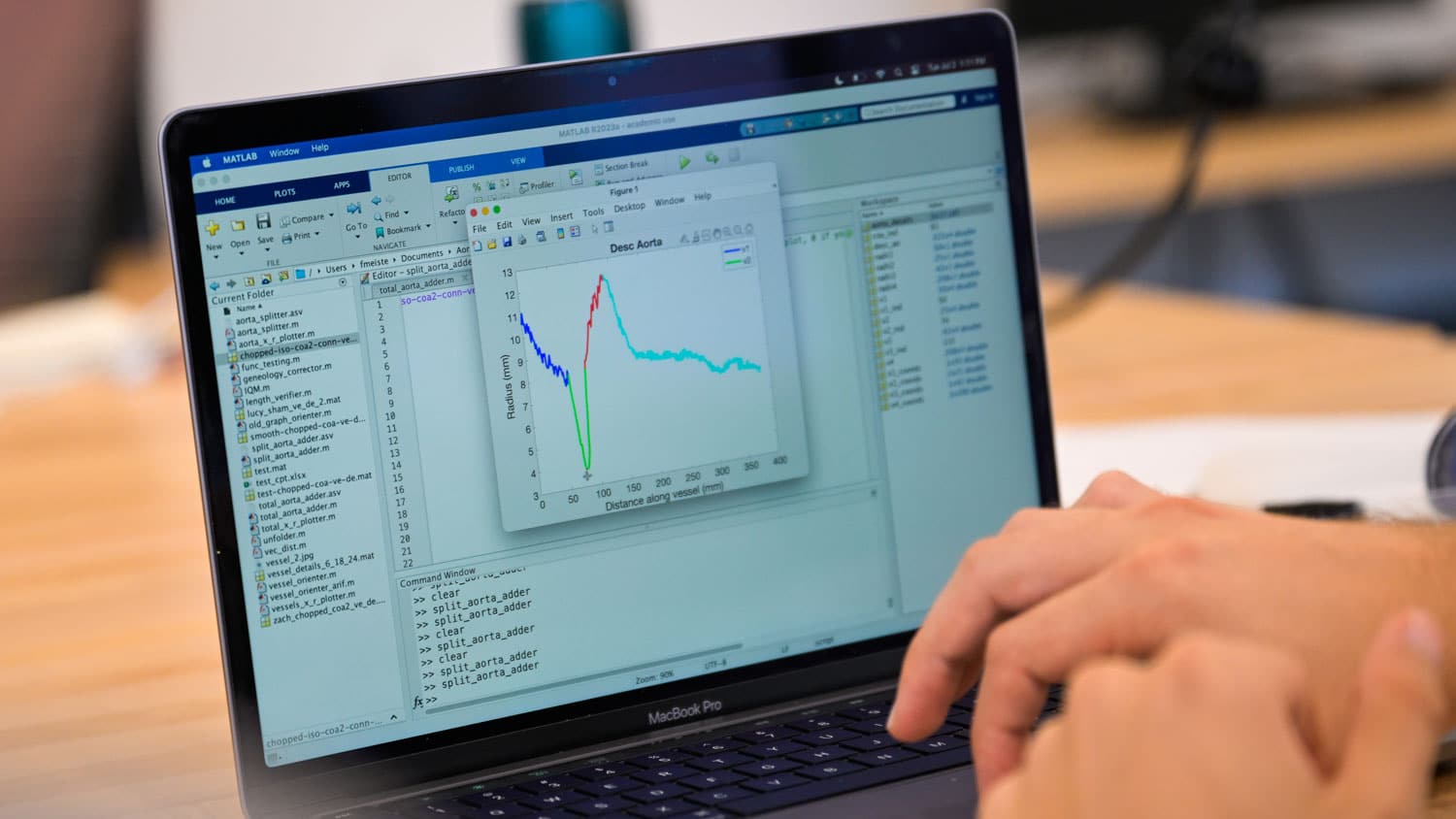
In mathematics professor Mette Olufsen’s lab, Ian Kriel worked with a team on fluid dynamics modeling using coding languages like MATLAB and C++ to predict the pressure and flow from an aortic coarctation system. These models can help determine the potential effects for someone with a specific type of coarctation, a heart defect typically present at birth in which the aorta is narrower than it should be.
The experience has informed Kriel’s plans for the future.
“Now that I’ve dabbled in coding, I found how useful it is for biochemistry and biology,” said Kriel, a biochemistry major at Christopher Newport University in Virginia. “And that’s changing the way that I’m thinking about my plans for the future. There’s a need for people who can code in biology, and that might be somewhere that I’m headed.”
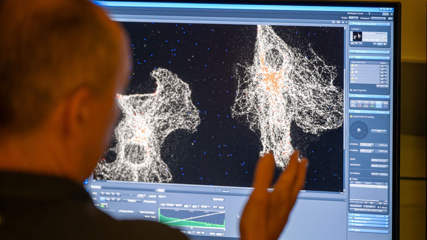
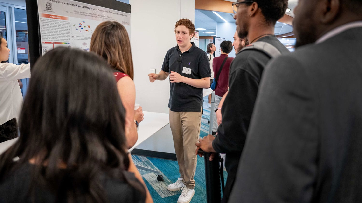
Meanwhile, Adam Nahor spent the summer working in the lab of David Muddiman, Jacob and Betty Belin Distinguished Professor of Chemistry, under the supervision of graduate students Sierra Hunter and Mary Wang.
The Muddiman group focuses on developing innovative mass spectrometry measurements to solve important biological problems. For instance, Hunter, Wang and Nahor used a laser to look at a zebrafish and find the molecular species found at each point. They were then able to form ion images showing the relative abundance of the molecules throughout the tissue.
Nahor, a pre-med engineering student at the University of Georgia, enjoyed doing research that combined biology and computation. His engineering background allowed him to approach the research in a different way.
“He actually took some computer science machine learning approaches to analyzing our samples, which really added a lot to our project,” said Wang.
Having Nahor in the lab over the summer also helped Wang and Hunter brush up on their chemistry knowledge and teaching skills.
“Teaching forces you to touch back on things you haven’t thought about in a while,” said Hunter. “If [Nahor] asked a specific question I didn’t know how to answer, I’d have to go back and figure it out.”
They Passed On Their Knowledge
The BioDynamics REU students put their understanding of microscopy to the test during an outreach activity with high school students. The students visited Catalyst, a program under The Science House, the College of Sciences’ K-12 outreach unit. Catalyst provides opportunities for high school students with disabilities to gain science, technology, engineering and mathematics (STEM) skills. The BioDynamics REU students split up into two groups, each one providing a presentation and hands-on activity on a different aspect of microscopy.
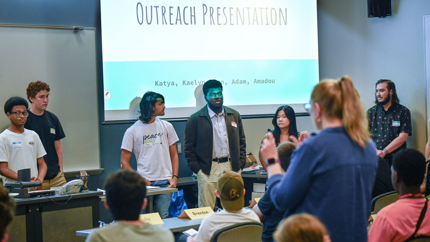
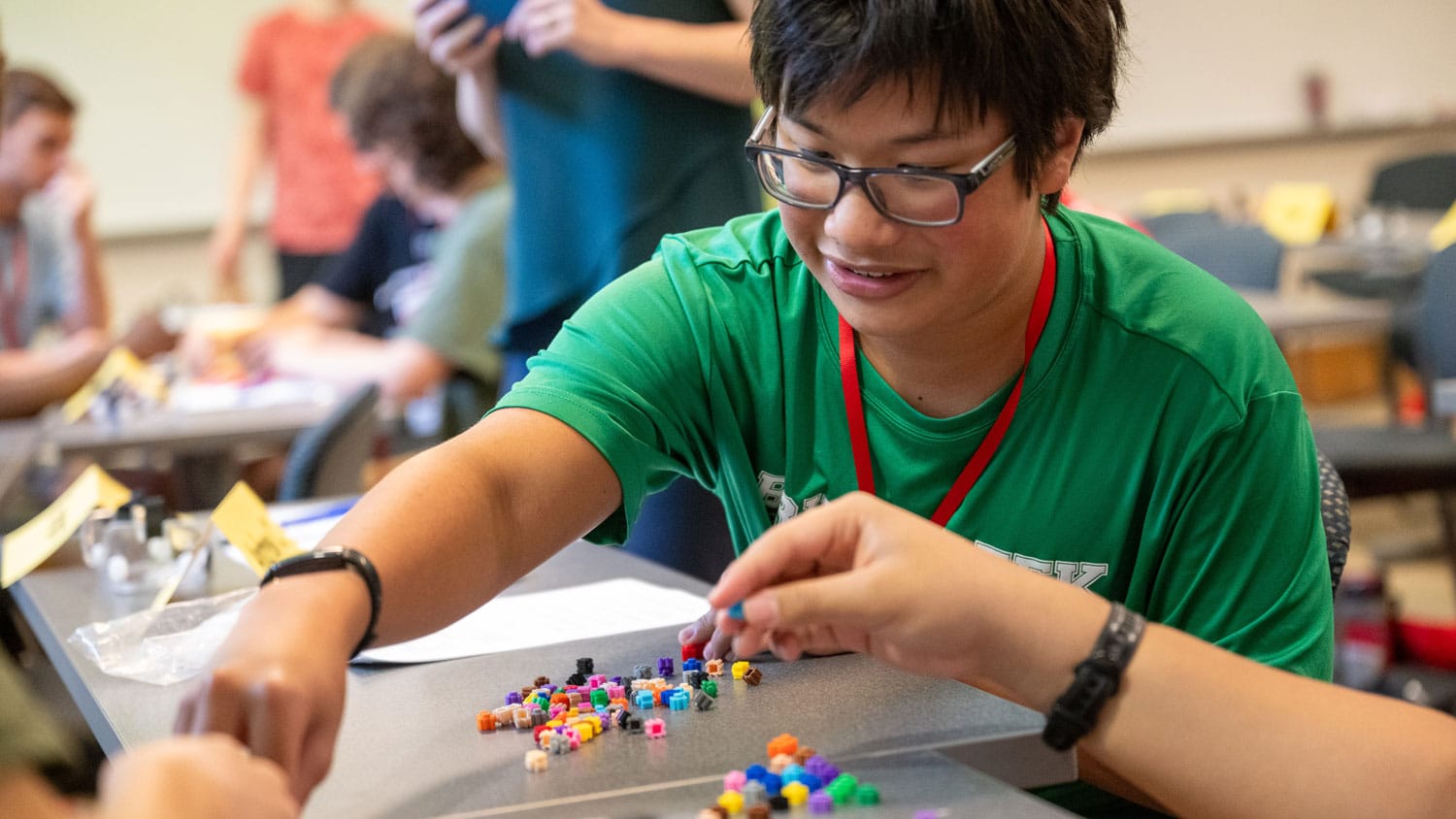
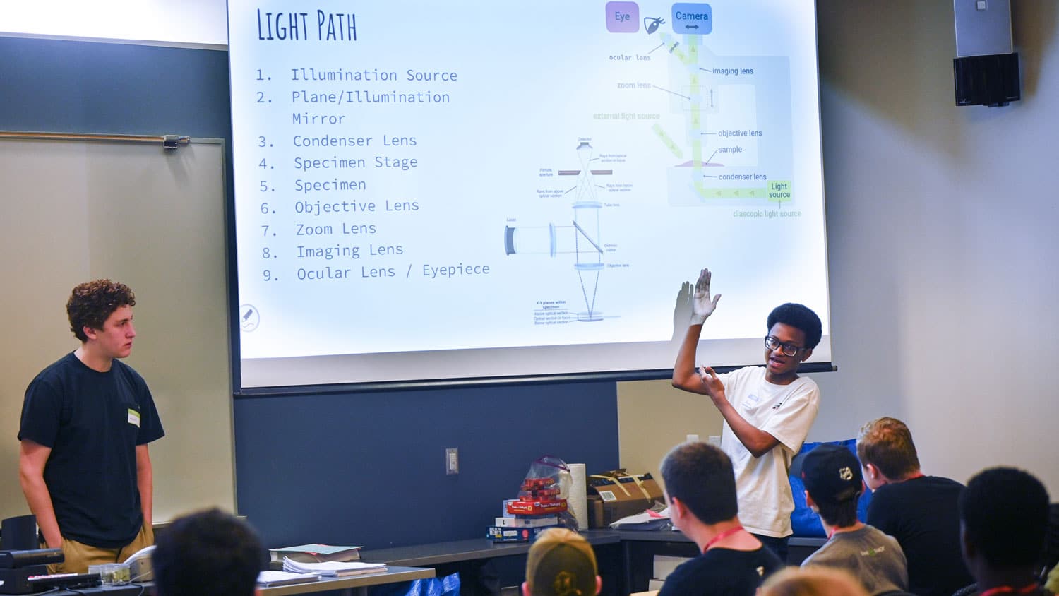
The Catalyst students learned about all the different parts of a microscope and how they all work together to allow scientists to study biological samples. They even built their own basic compound microscopes. Through other hands-on activities, they learned how to:
- Use a laser beam to manipulate light and enhance image focus and intensity.
- Increase image resolution with pixels.
- Use fluorescence to stain the parts of the cell they want to isolate.
- Prepare samples for viewing.
- Look at samples through various sizes of pinholes — which filter out unwanted light — to hone in on images.
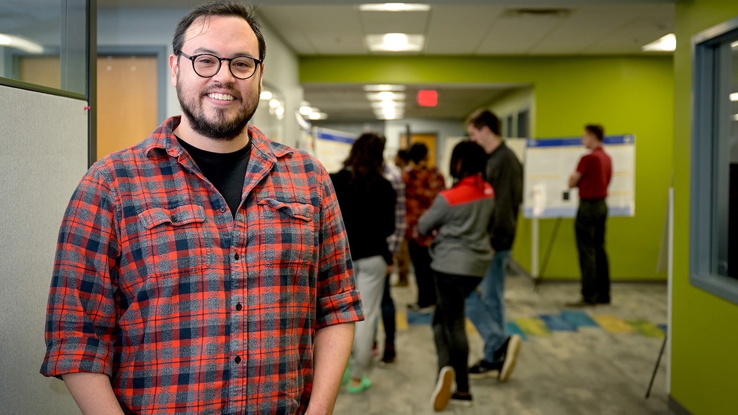
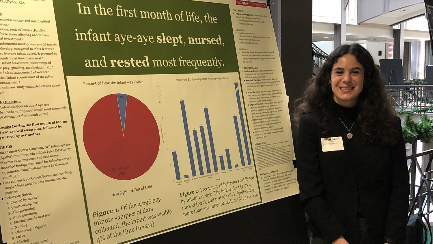
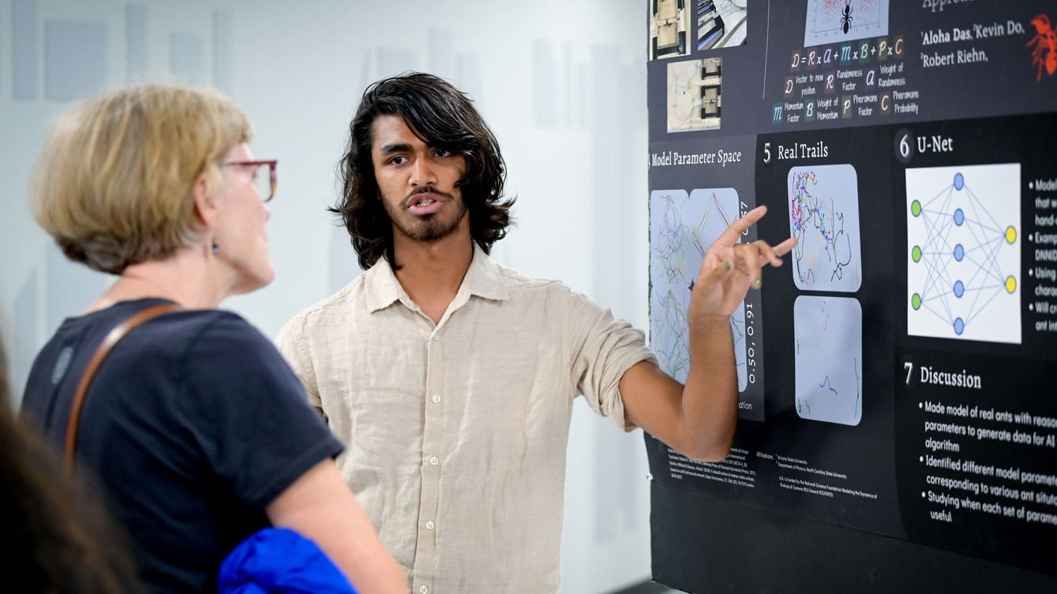
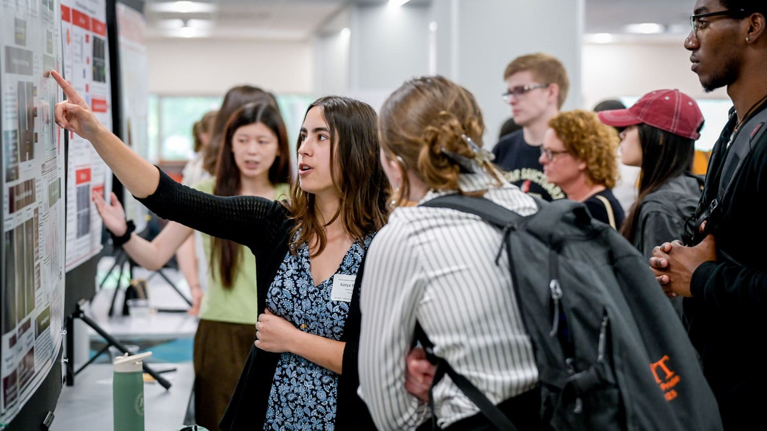
They Showcased Their Work
The BioDynamics REU students closed out their 10-week research experience by presenting their findings. On July 26, they joined student researchers from across NC State in setting up their posters at D.H. Hill Jr. Library for the Undergraduate Research Summer Symposium.
It was a summer to remember. Nothing makes us happier than equipping students — whether they’re from NC State or elsewhere — with the scientific knowledge and experience to take on the world’s most challenging issues. We were honored to host all the REU programs and can’t wait to do it again next year.
- Categories: